A Proposal for Research on Spinal Cord Repair Using Specific
Frequencies of Sound/Ultrasound
By Physicist Gary Wade
There is reason to believe that by the use
of specific frequencies of mechanical vibration (sound/ultrasound) that scar
tissue generated from spinal cord injury can be made to go embryonic-looking. Furthermore, that once these scar tissue
cells have become embryonic-like or looking (dedifferentiation) they can then
redifferentiate into the normal glia and other type
cells needed to repair the spinal cord.
The belief in the possibility of
converting scar tissue into embryonic like cells that will redifferentiate
into the needed normal cells for repair is based on several sources: 1) the work of Robert
Becker, MD, 2) the physical structures
of ion gates or ion channels on the various human cell types, and 3) my own work of converting fibroblast cells in
scar tissue into the type of cell(s) that should be there, if the scar tissue
were not there.
Dr. Robert O. Becker, MD, demonstrated
experimentally that both the salamander and mammals have the same tissue
regeneration mechanisms. These
regeneration mechanisms come into play after severe traumatic tissue injury (ref.
1). However, the mammal in general
compared to the salamander is deficient in the ability to send a sufficient
negative electric current to the damaged region, which manifests itself as hydroxyl ions
generation in the damaged tissue region (ref. 2). Becker demonstrated that if the mammal’s
ability to supply a negative electric current to a severely damaged region (
i.e. amputated leg) was artificially supplemented to the level comparable to
that used by a salamander, then the mammal (rat) would re-grow the amputated
leg (ref. 3). One of the necessities for
mammals to regenerate damaged tissue is for some cell types to dedifferentiate
into a embryonic-like cells and then redifferentiate into the needed cell types for tissue
repair or regeneration. In salamanders,
their red blood cells, which unlike those in mammals are nucleated, play this
role. In mammals Becker demonstrated
that fibroblast cells could be made to look embryonic and presumably can carry
out this same role in mammals.
In experiments carried out at the Center
for Complex Infectious Diseases (CCID) in
In field trials carried out with the same
type of magnetic pulse equipment used at CCID, we have been able to repair all manor and kind of fibroblast cell based scar tissue and
would be scar tissue damage in horses and humans. However, we have been unsuccessful with this equipment
use on spinal cord scar tissue, which is not fibroblast cell based. This does not mean that there might not be
some other pulsed magnetic field approach that would work to convert spinal
cord scar tissue to the needed normal spinal cord tissue structure.
Since, as Dr. Becker has shown, the
principles and mechanism of body repair and tissue regeneration for the
salamander and mammals seems to be essentially
identical, should we not expect a mammal to be able to repair or
regenerate from spinal cord injury, the same way a salamander does, if we just
artificially facilitate repair conditions?
One way to artificially facilitate repair conditions is to open up
specific ion gates on spinal cord scar tissue cells and change their cytoplasm
ion concentrations and have them become embryonic-like. Following from Becker’s work and my own work
with pulsed magnetic fields, it should be expected that the scar tissue cells
at the surface of the scar tissue, after they convert to embryonic like cells,
will redifferentiate to the type of cell that should
be there, if no scar tissue were there at the cells’ location. In studying fibroblast cell cultures exposed
to pulsed magnetic fields, it was only the fibroblast
cells at the edge of the cell culture that converted over to embryonic-looking
cells. The fibroblast cells in the bulk
of these cell cultures were butted up against each other and presumably
physically connected together. When
these cells were exposed to the magnetic pulses they showed some subtle
transient morphological changes but continued to look like fibroblast
cells. It was therefore found as
expected that several, to many repeated exposures of
the scar tissue, in field trials, were required to entirely convert the scar
tissue to normal tissue. The scar tissue
is a matrix of mainly fibroblast cells and collagen fibers. The fibroblast cells generate and maintain
the triple stranded electrically conductive protein collagen, which gives the
scar tissue its connective strength. As
the fibroblast cells are converted to normal tissue cells, they no longer
maintain the collagen matrix and in time it goes away leaving normal tissue
behind.
Through
the repeated (once per 24 hours) short exposure times (perhaps 5 to 15 minutes)
of spinal cord scar tissue cells to specific frequencies of sound/ultrasound
that open up specific ion gates, the scar tissue surface cells can possibly be
made to go embryonic-like and then turn into normal healthy neural tissue and
therefore spinal cord repair is accomplished.
With this possibility at hand it becomes a question of what are the
various mechanical vibration frequencies for the various metal ion gates and
other ion gates used by scar tissue cells.
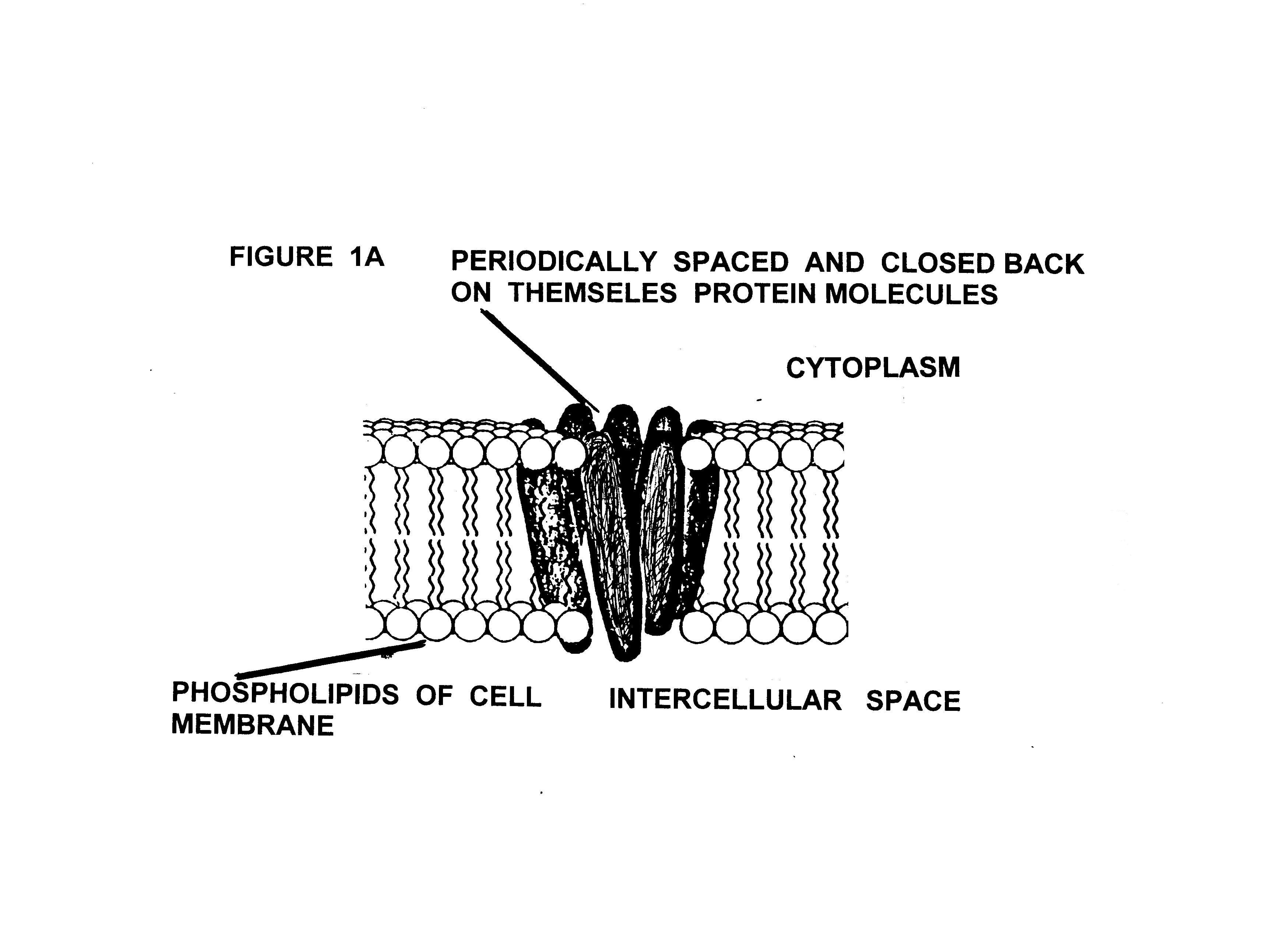
When looking in the literature at the
various now-known ion gate structures, a familiar
pattern appears. That pattern is that in
general the ion gates are made up of several nearly identical elongated trans bi-lippid membrane proteins
(see Figures 1A,
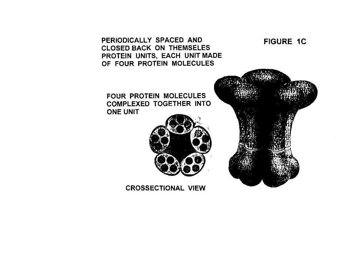
Figure 1B, and Figure 1C). Looking in a
direction at right angles to the cell membrane surface into the cell these
membrane crossing proteins form a circular closed-on-themselves pattern. In the middle of this circular protein
pattern, about half way through the cell bi-lipid membrane, the protein
molecules are very close together and they have amino acid sequences that only
allow the ion water complex for which the gate is for to be there. When the appropriate signal is received by
the ion gate, the center channel slightly enlarges or is unplugged and that ion
type streams into or out of the cell depending on which ion gate is
activated. Because these ion gate
structures in general, from a physics point of view, are essentially
periodically spaced masses which are elastically coupled together and form
a closed back-on-itself system, they will have specific mechanical vibration
frequencies with which they will go into resonant vibration. This is a known fact from the old German
mathematician eigenfunction problem/solution for the
standing waves on
a string with periodically spaced mass
beads of equal mass and with circular boundary conditions (closed back on
itself). What this means is, that if
these ion gate structures are exposed to their primary natural mechanical
resonance frequency, they can become open to ion transport for a significant
part of each resonant cycle. This is
because the opening up and closing down of the central ion channel cross sectional
area is part of the resonant mechanical vibration motion. In other words, the gates become very leaky
and are effectively open while exposed to their primary resonance
frequency. Because of the relatively
large masses of the ion gate proteins and the relatively low strength of
elastic coupling between gate proteins, the gate resonance frequencies should
be relatively low;
Perhaps as low as several hundred to several thousand cycles per
second.
At some university or other research
facility that has in operation labs that use ion type specific microscopic
probes for monitoring specific ion type
transport across cell membranes, a set of experiments needs to be
performed; Namely, monitoring a
specific cell’s ion transport of various ion types across the cell membrane as
a function of exposure to mechanical
vibration frequency. In our particular
case, we are
interested in studying the spinal cord scar tissue cells of both rats and
humans. Once the various gate
frequencies for the various metal and other ions are obtained, experiments can
be performed to find the frequency combinations, time of exposures, and
intensities to make the scar tissue cells in rats and humans go embryonic-
like, but without cell death and/or cell
damage. From here, a short period of
tests on rats with induced spinal cord scar tissue can be carried out. If the tests with rats are successful, then on to human trials. If the FDA is being consulted and puts up
its normal “let us wait for a few more years to test on humans” routine, immediately
do the human trial out of the country.
Note: There is no legitimate reason to put other species
of test animals in the hundreds to thousands, into misery doing these spinal
cord tests. Any research institution
that thinks mass animal tests are required, should not
be involved in this research.
References:
1) The Body Electric,
Electromagnetism and the Foundation of Life, by Robert O. Becker, M.D., and
Gary Selden, and illustrated by David Bichell; ISBN 0-688-06971-1
2) A Physicist’s View of the Use
of Feeble Electric Direct Currents to Repair Tissue and Replace Body Parts
(Part One), by Gary Wade, Health Freedom News, February 1996, Pages 22 to 33. (see attached article).
3) The Body Electric, Pages 152
to 155.
4) The DNA Helix and How It Is
Read, Scientific American, December 1983, Vol. 249, No, 6, pp. 94-111.
Note to would-be
researchers:
In my experiments with sound/ultrasound
generation for experimentation on microbes and cell cultures, I have used 1/8th
pie cut pieces of the standard 2 inch in diameter and 1/10 inch thick circular
piezoelectric transducer elements that are used in ultrasonic cleaner
tanks. These 1/8th pie cut pieces are
easily ran with the standard off the shelf electronic tech signal function
generator found In physics labs and used by electronic techs. I usually epoxy the 1/8th cut element to a
small metal rectangular piece and then clamp or fixture the metal plate to the
microscope stage or slide holder or side of dish. The standard piezoelectric elements are
polarized such that one side of the transducer element should always be
maintained at a positive potential to the other side, so that the element does
not degrade in its ability to generate mechanical vibrations with applied
electrical voltage oscillations. If
higher amplitude of mechanical vibration is desired or required, the signal
from the signal function generator can be amplified by using a linear
amplifier. For example, if the frequency
range of interest is from say 20 cycles per sound (cps) to 30,000 cps then a
good audio amplifier will do the job.
This frequency range is expected to be the range in which the various
ion gates on cell membranes will have their resonance frequencies. The piezoelectric element only transforms
voltage sign waves into pressure sine waves.
Keep in mind that one side of the
piezoelectric element needs to be free to expand and contract in the air or
liquid environment being used for experimentation.
For the frequency range above, standard
off the shelf speakers can be used.
However, some kind of sound enclosure will be needed so as not to bug
everyone to tears.
IF YOU FOUND THIS ARTICLE OF REAL VALUE, PLEASE MAKE A HARD COPY WHILE STILL AVAILABLE.